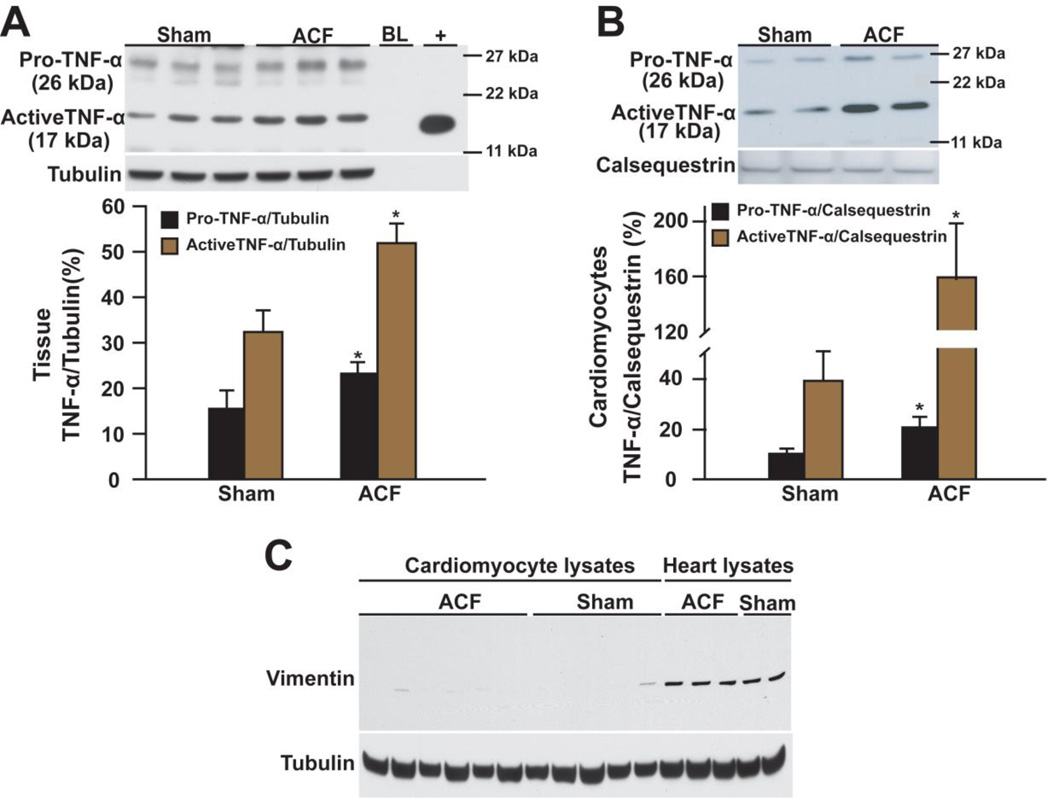
Figure 4. Western blot of TNF-α in LV tissue and isolated cardiomyocytes of 24 hour ACF.
Pro- and active TNF-α in (A) heart lysates and (B) cardiomyocytes from ACF vs. shams. “BL” loading dye. “+” recombinant active rat TNF-α. Densitometric analysis is presented below. *P < 0.05, **P < 0.01 vs. sham. (C) Western blot of vimentin in cardiomyocytes and heart lysates (positive for vimentin) to ensure the purity of cardiomyocytes. Tubulin or calsequestrin was used as loading control.

