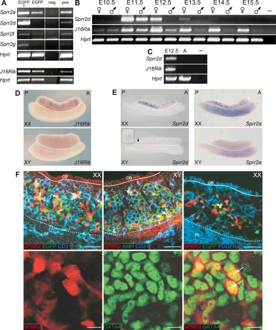
Figure 1. Expression analysis of Sprr2d and 1700106J16Rik in developing gonads.
Semi-quantitative RT-PCR analysis (A–C). (A) Expression of Sprr2a, d, f, g and 1700106J16Rik (J16Rik) in FACS-sorted cells from E13.5 B6 Sry-EGFP transgenic ovaries. Each gene was more highly expressed in EGFP+/CD31- cells (EGFP+) than in EGFP-/CD31- cells (EGFP-). (B) Analysis of Sprr2d and J16Rik expression in E10.5 urogenital ridges and E11.5–E15.5 gonads. In XX gonads (♀), Sprr2d expression was highest at E11.5 and rapidly decreased thereafter. In XY gonads (♂), Sprr2d was detected only at E11.5. J16Rik was expressed at very low levels in both sexes at E10.5, and expression increased at E11.5 in both sexes and was maintained in XX gonads until at least E15.5. In XY gonads, J16Rik expression decreased after E11.5, and was essentially undetectable by E13.5. Both genes were expressed more highly in XX than in XY gonads at each developmental stage analyzed. (C) Neither gene was expressed in adult ovaries. In (A–C) a reaction was performed without template as a negative control (neg or –), and Hprt served as the endogenous control. In (A) additional positive controls (pos) were performed using RNA from non-sorted cells from dissociated E13.5 B6 Sry-EGFP transgenic ovaries for the Sprr2 panel, or using RNA from intact E13.5 B6 ovaries for the J16Rik panel. In (C) E12.5 XX gonads were used as a positive control (leftmost lane). WISH analysis (D and E). (D) In E13.5 gonad/mesonephros complexes, J16Rik expression was detected in a distinct subset of cells within XX gonads, but not in XY gonads or mesonephroi of either sex. (E) In E12.5 XX gonads, Sprr2d and Sprr2a were expressed in discrete cells with a posterior bias. In E12.5 XY gonads, Sprr2d was expressed in just a few cells at the very posterior end (arrow and inset) and Sprr2a expression was not detected. A, anterior; P, posterior. Gonads are above the dotted line and mesonephroi are below. (F) Localization of SPRR2 protein(s) was analyzed using WIHC. E12.5 B6 Sry-EGFP transgenic XX gonads were labeled with antibodies to SPRR2 (red), EGFP (green) and CD31 (blue) (upper left panel). CD31 marks germ and endothelial cells and Sry-EGFP marks pre-granulosa cells. SPRR2 was expressed in a subset of CD31-negative somatic cells, some of which also expressed EGFP (yellow and arrows). E12.5 B6 XY gonads were labeled with antibodies to SPRR2 (red), AMH (green) and CD31 (blue) (upper middle panel). AMH marks Sertoli cells. SPRR2 was not expressed in CD31-positive germ cells or endothelial cells but was expressed in a subset of AMH-positive Sertoli cells (yellow and arrows). E12.5 B6 Sry-EGFP transgenic XX gonads were labeled with antibodies to SPRR2 (red) and PDGFRα (blue) (upper right panel). Sry-EGFP transgene expression (autofluorescence) is green. EGFP expressing pre-granulosa cells and SPRR2 expressing cells were located in the innermost regions of the gonad, and EGFP and SPRR2 expression overlapped in some cells (yellow and arrow). PDGFRα marks coelomic epithelium and interstitial somatic cells, and its expression did not overlap with either EGFP or SPRR2. In each upper row image, the coelomic epithelium (ce) is located above the solid line, and the mesonephros (m) below the dotted line. In the lower row, E13.25 XX gonads were labeled with antibodies to SPRR2 (red) and GATA4 (green). GATA4 marks somatic cell nuclei. SPRR2 was expressed in a subset of GATA4-positive cells, and SPRR2 protein was co-localized with GATA4 in cell nuclei (lower right panel, yellow and arrows). SPRR2 also was located in the cytoplasm. Scale bars: 50 μm (upper row), 10 μm (lower row).

