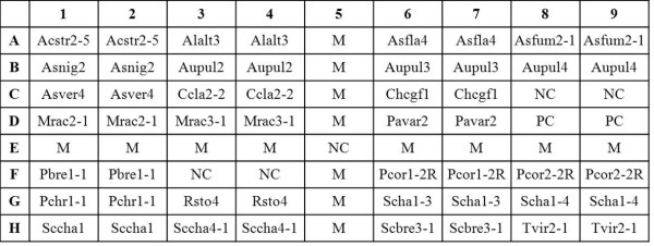
Figure 1.
Layout of oligonucleotide probes on the array (0.8 × 0.7 cm, 9 by 8 dots). The probe "PC" was a positive control and the probe "NC" was a negative control (tracking dye only). The probe "M", a position marker, was an irrelevant probe labeled with a digoxigenin molecule at the 5' end. The corresponding species names and sequences of all probes are listed in Table 2. All probes used for fungal identification were spotted on the array in duplicate.

