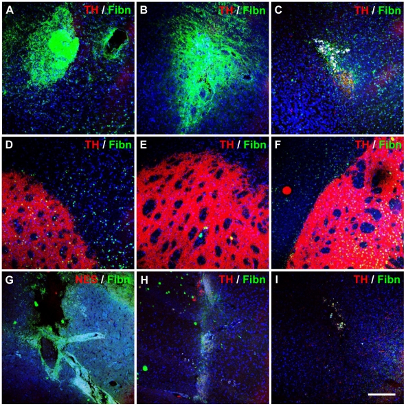
Figure 9. Matrix deposition and lack of dopaminergic neuronal differentiation after transplanting neuronal-primed hMSCs and OECs.
Fibronectin and TH expression were assessed in striatal and nigral grafts at 1-day (left column), 7-days (middle column) and 21-days (right column). Representative images are shown of (A-C) neuronal-primed hMSC graft sites in the striatum, (D–F) corresponding regions in the contralateral hemisphere, and (G–I) injection sites in sham controls (n = 3). Immunohistological analysis revealed a fibronectin-positive matrix surrounding hMSC graft sites from 1-day post-transplantation (A and B), however, this was markedly reduced by 21-days (C). The concomitant loss of transplanted cells suggests that the fibronectin matrix was deposited by the graft. Furthermore, sham injected animals did not exhibit dense fibronectin expression (G–I). TH expression was not detected in any graft sites or sham-injected sites, and was only expressed in the unlesioned hemisphere (D–F). Similar results were observed in OEC co-transplanted recipients. Images shown are maximum projection z-stacks. Nuclei were counterstained with DAPI (blue). Scale bar: 150 µm.

