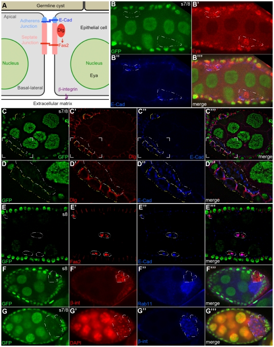
Figure 5. rab11-null follicle cells lose their polarity, delaminate from the epithelium and invade the neighboring germline cyst.
(A) Schematic diagram of follicle epithelial cell polarity. Markers used in this study are highlighted [adapted from [13]]. (B–G″) Confocal images of mosaic stages 7/8 egg chambers 4–6 days ACI. The rab11-null clones are marked by their absence of GFP expression and are outlined with dashed lines. (B-B′″) nGFP (green), Eya (red), E-cad (blue). All of the rab11-null cells stain positive for Eya, consistent with an epithelial cell fate. In contrast to the strict apical expression pattern of E-cad in neighboring wildtype cells, the protein is highly enriched in intracellular compartments in the rab11-null cells (also see D″ and E″). (C-C′″) nGFP (green), Discs large (Dlg) (red), E-cad (blue). (D-D′″) Enlarged views of the bracketed regions shown in (C-C′″). Note that rab11-null cells that are still embedded in the epithelium (outlined in yellow) exhibit wildtype or near wildtype (basolateral) expression patterns for Dlg and mostly normal (mostly apical) expression pattern for E-cad. In contrast, the rab11-null cells that have delaminated from the epithelium (outlined in white) exhibit a vesicular staining pattern for E-cad, while Dlg is dispersed throughout the cell and /or completely absent. (E-E′″) nGFP (green), Fas2 (red), E-cad (blue). Three clusters of delaminated rab11-null cells are outlined. Each cluster contains two cells. None of the cells exhibit apical-basal polarity as evident by the vesicular-like staining pattern of both Fas2 and E-cad. (F-F′″) nGFP (green), ß-integrin (ß-int) (red), Rab11 (blue). Note, the donut-shape distribution pattern of ß-int, which suggest that the some of the protein is still on the cell surface. All of the other examined cell surface markers exhibit a strictly intracellular staining pattern in delaminated cells. The circled rab11-null clone, along with the one shown in (G) is situated in a bubble between the epithelium proper and the germline cyst, which partially accounts for the weak ß-int signal in the flanking wildtype epithelial cells. Nevertheless, the ß-int signal was reproducibly more intense in the rab11-null epithelial cells than in wildtype epithelial cells. (G-G′″) nGFP (green), DAPI (red), ß-int (blue). A large (>50 cells) rab11-null clone in the posterior portion of the egg chamber is circled. Note that the ß –int staining pattern in this clone is more vesicular in nature than that in the previous panels. Most other similarly large clones were also located in the posterior portion of the egg chamber and like the one shown wedged between the follicle cell epithelium and the oocyte.

