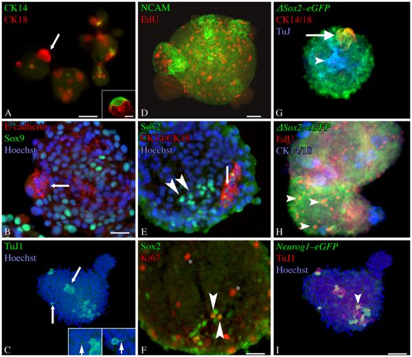
Figure 2. Olfactory neurospheres express cell-type specific markers characteristic of intact olfactory epithelium.
ONS were stained as whole mounts (A-D) or cryosectioned and stained as described (E–F). A) Occasional CK14(+) cells can be found in ONS, but they appear with less frequency than any other differentiated cell type examined. Some of the CK14(+) cells have morphologies highly reminiscent of the in vivo HBC counterparts (inset). CK18(+) cells are also present in the spheres and are distinct from CK14(+) cells. These often occur in subdomains that appear as “buds” of morphologically homogeneous cells (arrow). B) Staining for E-cadherin, a marker of sus and Bowman's Duct and Gland structures in vivo, also labels these subdomains (arrow). Numerous Sox9(+) cells, a marker of Bowman's Duct and Gland cells in the OE, are present in ONSs. C) TuJ-1(+) cells are found throughout ONSs. Often, these cells have processes (arrows–shown at higher power in insets) that resemble axons (left inset) or apical dendrites (right inset). D) NCAM(+) also labels a neuronal population present in ONS. EdU(+) cells indicate that DNA synthesis is actively occuring in the ONSs. E) Sox2(+)/CK14(−)/CK18(−) cells (arrowheads) and Sox2(+)/CK14(+)/CK18(+) cells (arrow) are found in ONSs. F) A number of Sox2(+) cells are Ki67(+) (arrowheads). Additional cells in the ONSs are also Ki67(+) (asterisk). G, H) ONSs derived from ΔSox2eGFP neonates The vast majority of cells in the spheres are GFP(+). These include both cytokeratin 14/18(+) cells (arrow) found in subdomains and TuJ-1(+) cells stretching throughout the ONS (arrowhead). H) Many of the GFP(+) cells are also EdU(+) and cytokeratin 14/18 (−) (arrowheads). These criteria define GBCs in vivo and indicate their presence in ONSs. Separate CK14 and CK18 antibodies were visualized with secondary antibodies conjugated to the same flourophore to allow for detection of additional antigens (TuJ in G, EdU in H). I) ONSs derived from Neurog1eGFP neonates. A number of cells in the spheres are GFP(+) and many of these are TuJ1 (+) and/or PGP9.5(+) (arrowheads). Scale bar in A is 50 μm. Scale bar in the inset of A is 10 μm. Scale bar in B is 25 μm and applies to B and E. Scale bar in D is 50 μm and applies to C and D. Scale bar in F is 25 μm. Scale bar in I is 50 μm and applies to G and H.

