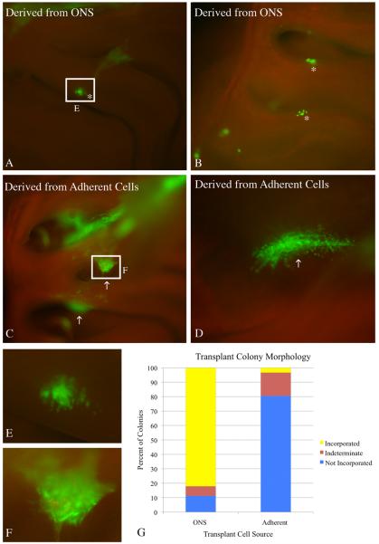
Figure 4. ONS-derived cells engraft appropriately into the lesioned recovering epithelium.
Cells derived from 8 DIV ONS were compared to cells derived from 8 DIV adherent cultures in a transplantation assay that tests engraftment capacity. A, B) 2 weeks after transplantation, cells derived from ONS cultures formed primarily smaller colonies that have incorporated and integrated into the regenerating epithelium (asterisks). C, D) In contrast, cells derived from adherent cultures formed primarily large, sprawling colonies (arrows) that have not incorporated into the OE of methyl bromide-lesioned host animals. The colonies appear to sit above the epithelium or bridge adjacent areas of epithelium via synechiae. E) Inset of boxed region in A shows more detailed morphology of an ONS–derived colony. The GFP(+) cells intercalate extensively with host cells as seen by the punctate GFP signal intermixed with GFP(−) host cells, suggesting appropriate incorporation into the epithelium. F) Inset of boxed region in C shows more detailed morphology of an adherent culture–derived colony. The GFP(+) cells do not intercalate extensively with host cells. G) Engrafted colonies from three host animals for each transplant condition were classified into three categories: (1) incorporated (resembling the colonies identified by asterisks) – depicted in yellow; (2) not incorporated (resembling the colonies identified by arrows) – depicted in blue; or (3) undefined (unable to definitively classify) – depicted in plum. The graph clearly shows that colonies resulting from ONS-derived cells result in a higher percentage of incorporated colonies. Yates Chi-square analysis of the raw data indicates a significant difference between the two conditions (Yates Chi-square = 36.999, Yates p-value = 1 × 10⁻⁸ ).

