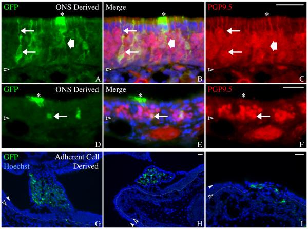
Figure 5. ONS-derived colonies contain cell types that resemble adjacent host OE.
A-F) Coronal sections of ONS-derived colonies confirm whole mount observations that the sphere-derived cells intergrate into the regenerating epithelium. Neurons (arrows) and sustentacular cells (asterisks) are identified based on morphology and immunostaining. GFP(+)/PGP9.5(+) neurons with an apical dendrite (upper arrow in A-C) are seen in the transplant-derived colonies. Transplant-derived sustentacular cells (asterisks) are identified as GFP(+)/PGP9.5(−) cells with apically positioned nuclei and footprocesses extending toward the basal lamina (wide arrow). Scale bar in C is 25 μm and applies to A-C. Scale bar in F is 25 μm and applies to D-F. G-I) Coronal sections of adherent culture–derived colonies confirm whole mount observations that these cells do not incorporate appropriately into regenerating epithelium. Endogenous GFP was compared with Hoechst labeling of all nuclei. The colonies resulting from adherent culture–derived cells appear spindle-shaped and mesenchymal and thus, are distinct morphologically from the normal differentiated cell types found in the OE. The basal lamina is designated by a black arrowhead and thin dotted line, the apical surface of the epithelium is designated by a white arrowhead. Scale bar in H is 25 μm and applies to G. Scale bar in I is 25 μm.

