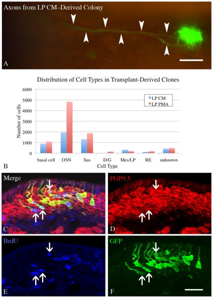
Figure 7. Colonies derived from spheres cultured in LP PMA have an increased number of neurons.
ONS derived from constitutively expressing GFP donor neonates were cultured in LP CM or LP PMA for 8 DIV, then dissociated and transplanted into 1 day post methyl bromide-lesioned host animals. Animals were sacrificed 2 weeks after transplantation and screened for GFP(+) donor-derived colonies. A) Large colonies were seen in both conditions (LP CM shown here), with some having visible axons projecting back towards the olfactory bulb (arrow). Scale bar is 100 μm. B) All colonies from LP CM and LP PMA hosts (n=3 per condition), were counted and the cell types present were documented. Chi–square analysis indicates that the two distributions are significantly different from one another (Chi–square = 681, p < 0.0001) and neurons significantly contribute to this difference (Chi–square = 459.9, p < 0.001). C-F) A cohort of host mice were pulsed with 30 mg/kg BrdU 8 days after transplantation to label cells in S-phase and then allowed to survive for an additional 6 days (the chase phase) before fixation. Many neurons arise in vivo from transplant-derived cells (arrows) as shown by co-labeling with GFP to identify donor–derived cells, PGP9.5 to identify neurons, and BrdU to identify cells labeled during the pulse. Scale bar in F is 25 μm applies to C-F.

