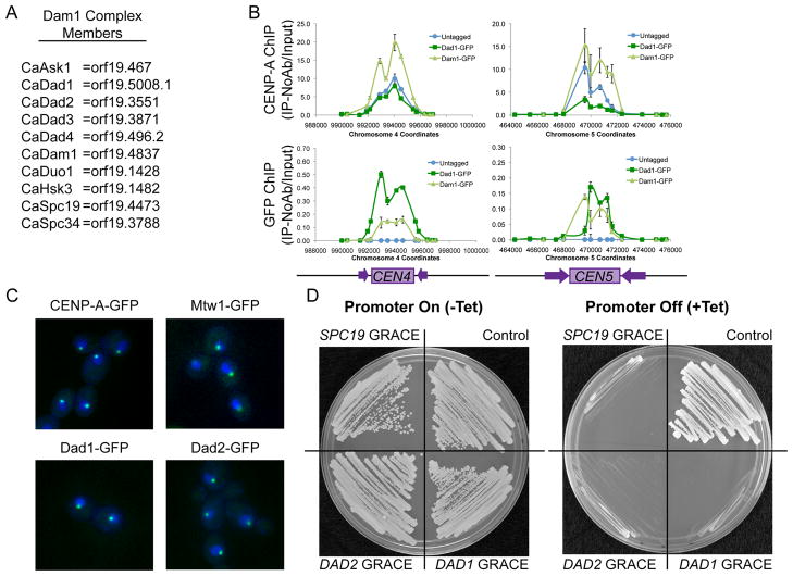
Figure 1. C. albicans Dam1 complex proteins bind centromere DNA regions and are essential in C. albicans.
(A) C. albicans Dam1 complex homologs identified by reciprocal protein-protein BLAST and Pfam family analysis (see also Table 1S). (B) ChIP with anti-CENP-A (upper panel) and anti-GFP antibodies (lower panel) at CEN4 and CEN5 DNA regions in BWP17 (untagged, blue), Dad1-GFP (dark green), and Dam1-GFP (light green) strains shows co-localization of CENP-A and Dam1 complex members on centromeric DNA. Data shown are mean ± SEM of qPCR analysis of each primer set in duplicate and are representative of 3 biological replicates. (C) GFP-tagged CENP-A, Mtw1-GFP, Dad1-GFP and Dad2-GFP (green) show similar localization patterns within DAPI-stained nuclei (blue). Cells were imaged by fluorescence microscopy at 1000x total magnification. (D) GRACE (Gene Replacement and Conditional Expression) DAD1, DAD2, or SPC19 strains and the control strain (SC5314) all exhibited robust growth on SDC-Ura media (left panel), but failed to grow when the Tet-off promoter was repressed by the addition of 50μg/ml tetracycline to SDC-Ura media (right panel).

