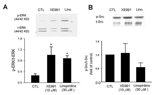
Figure 6. Blocking KCNQ2/3 selectively increased ERK1/2 phsophorylation.
Western blot analysis was performed to inspect the total and phosphorylated protein levels of several intracellular signaling molecules in ES cell-derived neural progenitor cells. Both XE991 and linopirdine (5 day exposure) enhanced the levels of phsophorylated ERK1/2 (pERK). The bar graph shows that, due to the increased pERK1/2 and/or decreased total ERK (tERK), the ratio of pERK versus tERK was significantly increased. Meanwhile, Src phosphorylation was not significantly changed. N = 5; *. P < 0.05 vs. controls.

