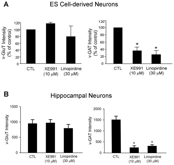
Figure 8. Blockage of KCNQ2/3 channel affected synapse formation.
Bar graphs summarized fluorescent intensity of v-GluT and v-GAT immunostaining in ES cell-derived neuronal cells and hippocampal neurons. The expressions of v-GluT and v-GAT were used as markers for excitatory and inhibitory synapses, respectively. A. Quantification of intensities of v-GluT and v-GAT staining in mouse stem cell-derived neurons with and without XE-991 or linopirdine for 5 days after neuronal induction. B. Quantified data from experiments with hippocampal neurons of 14-DIV. XE991 and linopirdine significantly reduced v-GAT expression without affecting v-GluT expression. Mean ± S.E. M. N ≥ 3 independent experiments. * P < 0.05 vs. vehicle controls.

