
Figure 1. L-DOPA- and D1R-mediated priming and post-test AIMs.
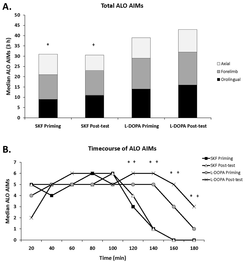
(A) Rats that received unilateral 6-OHDA lesions of the MFB were primed 3 weeks later with L-DOPA (12 mg/kg + benserazide, 15 mg/kg, sc; Experiments 1 and 3; n = 30) or the D1R agonist SKF81297 (SKF; 0.8 mg/kg,sc; Experiment 2; n = 8) and exhibited summed axial, forelimb, and orolingual (ALO) AIMs ≥ 25 by the last day of priming (3 h total). Following microdialysis test days, rats were given a post-test of L-DOPA or SKF alone in order to ensure AIMs stability over the course of testing. The distribution of axial (white), forelimb (gray), and orolingual (black) AIMs are represented. (B) Timecourse of AIMs for the last day of priming and the post-test of 6-OHDA-lesioned animals treated with L-DOPA or SKF. Differences in AIMs (expressed as medians) were analyzed with non-parametric Kruskal-Wallis tests and Mann-Whitney post hoc comparisons.
* p < 0.05 for SKF priming vs L-DOPA priming
+ p < 0.05 for SKF post-test vs L-DOPA post-test