
Figure 2. Effects of 6-OHDA-lesion, L-DOPA priming, and D1R agonist priming on basal extracellular striatal glutamate levels.
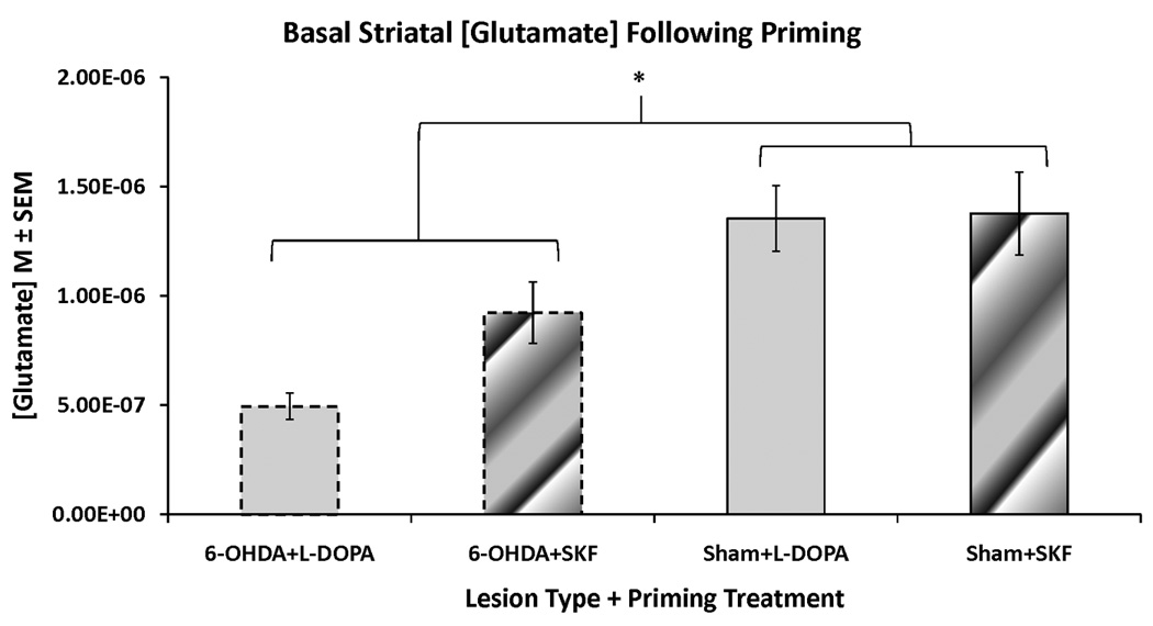
Rats in the first and second experiments received unilateral 6-OHDA or sham lesions of the MFB and 3 weeks later were primed with L-DOPA (12 mg/kg + benserazide, 15 mg/kg, sc) or the D1R agonist SKF81297 (SKF; 0.8 mg/kg,sc). Following priming, rats underwent a microdialysis procedure including 40 min of baseline sampling prior to any vehicle or drug injection. Striatal glutamate concentrations (M) from these baseline samples were averaged and analyzed with a two-way ANOVA (lesion group × priming treatment). The groups include: 6-OHDA+L-DOPA (n=12), 6-OHDA+SKF (n=9), Sham+L-DOPA (n=11), and Sham+SKF (n=7).
* p < 0.05 for 6-OHDA-lesioned groups vs sham-lesioned groups