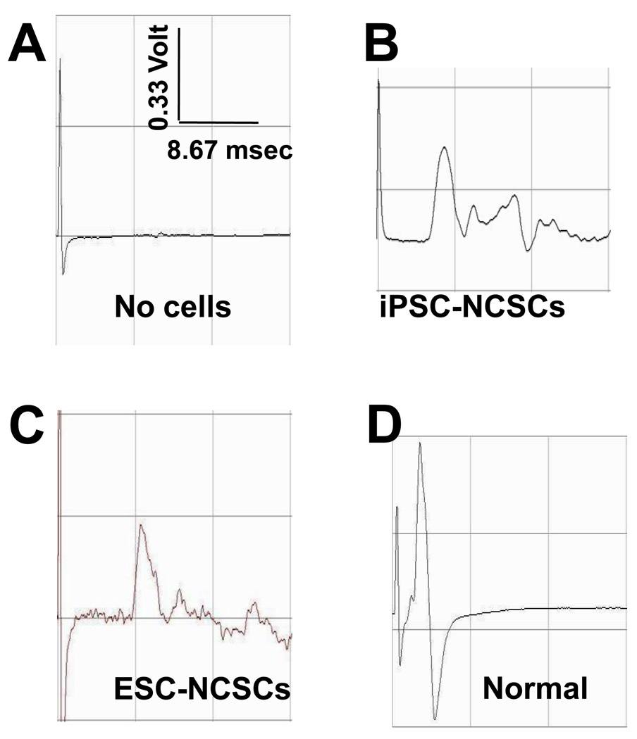
Figure 6.
Sciatic nerve regeneration by transplanting iPSC-derived NCSCs in nanofibrous nerve conduits. CMAP was measured at 1-month after surgery. Representative data are shown for the control group (without cells) (A), the group with iPSC-NCSC transplantation (B), the group with ESC-NCSC transplantation (C) and contralateral normal nerve group (D).

