
Figure 1.
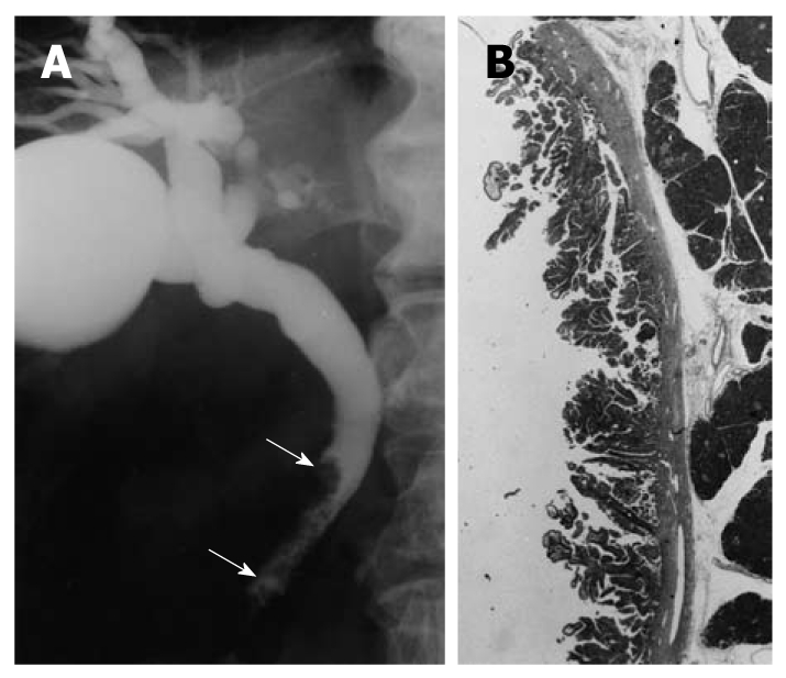
Cholangiographic finding of extrahepatic bile duct carcinoma in the nonicteric stage. A: Cholangiography shows a papillary tumor at the distal common bile duct (arrows); B: The histologic findings of the resected specimen showed papillary adenocarcinoma confined to the mucosal layer (hematoxylin and eosin, × 1).