
Fig. 2.
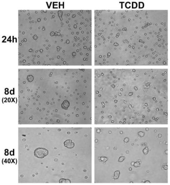
Morphology of differentiating SCp2 cells exposed to TCDD. Photomicrographs of SCp2 cells, induced to differentiate and exposed to vehicle (0.1% DMSO) or TCDD (1 nM), were taken at 24h and 8d after plating. Reduced alveolar-like structure formation is observed after TCDD exposure. Images of the 8d cultures were obtained using both low (20X) and high (40X) magnification objective lenses. h, hours; d, days.