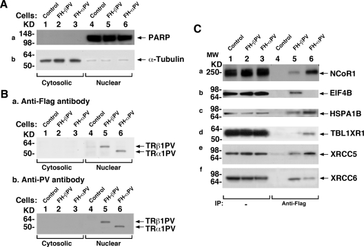
Fig. 2.
Physical interaction of NCoR1 with TR mutant isoforms in FH-βPV and FH-αPV cells. A, FH (controls) (lanes 1 and 4), FH-βPV (lanes 2 and 5), and FH-αPV (lanes 3 and 6) were cultured as described in Materials and Methods. Nuclear and cytoplasmic fractions were analyzed by Western blotting for the expression of markers for nuclear (PARP) (a) and cytoplasmic (α-tubulin) (b) fractions. (B-a) TRβ1PV and TRα1PV proteins were detected by monoclonal anti-Flag antibody, M2 (0.5 μg/ml), or by monoclonal anti-PV-specific antibody (2 μg/ml). B-b, Lanes 1 and 4 were from control cells as negative controls, indicating the specific bands detected in lanes 5 and 6. C, Association of NCoR1 and other nuclear proteins with TR mutants in FH-βPV and FH-αPV cells shown by coimmunoprecipitation. Nuclear extracts (1 mg) were immunoprecipitated with mouse anti-Flag M2 affinity gel followed by Western blot analysis with anti-NCoR1 antibody (C-a), anti-EIF4B antibody (C-b), anti-HSPA1B antibody (C-c), anti-TBL1XR1 antibody (C-d), anti-XRCC5 antibody (C-e), or anti-XRCC6 antibody (C-f). Lanes 5 and 6 were from nuclear extracts of FH-βPV and FH-αPV, respectively. Direct Western blot analysis (25 μg of nuclear extracts) is shown in lanes 1–3 as marked. IP, Immunoprecipitation.

