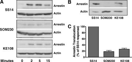
Fig. 5.
Agonist-induced membrane translocation of β-arrestin-2. CHO cells were transiently transfected with FLAG-β-arrestin-2 and sst2A receptor. After 48 h, cells were incubated at 37 C with 100 nm SS14, 1 μm SOM230, or 1 μm KE108 for the times shown. After cell lysis, membranes were purified by centrifugation as described in Materials and Methods, and the membrane fraction was subjected to SDS-PAGE. Membrane-associated β-arrestin-2 and actin were detected by incubating each blot with anti-FLAG and antiactin antibodies. Band intensities were quantitated from scanned autoradiograms using Scion Image (version 1.63). A, Time course of arrestin translocation to membranes after peptide treatment in representative experiments. The actin band provides a loading control. B, Direct comparison of membrane-associated β-arrestin-2 after treatment of cells with 100 nm SS14, 1 μm SOM230, or 1 μm KE108 for 5 min in a representative experiment. C, Mean ± sem (n = 6) for membrane-bound arrestin after treatment of cells for 5 min with different analogs in experiments like the one shown in B. The ratio of arrestin to actin was calculated in each sample and then expressed as a percentage of the arrestin/actin ratio observed in samples treated with SS14 for 5 min.

