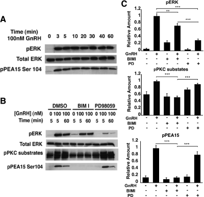
Fig. 1.
GnRH induces PEA-15 phosphorylation via PKC in LβT2 cells. A, Time course of GnRH-stimulated ERK and PEA-15 activation in LβT2 cells. Total ERK was used as a loading control. LβT2 cells were serum starved overnight before treatment with 100 nm GnRH for the indicated periods of time. Whole-cell lysates were subjected to a Western blot analysis using phospho-ERK- and phospho-PEA-15 (Ser104)-specific antibodies. Total ERK was used as a loading control. B, Effect of pharmacological inhibitors on GnRH-induced ERK and PEA-15 activation. LβT2 cells were serum starved overnight, pretreated with the following inhibitors for 30 min: 10 μm BIMI or 50 μm PD98059 and stimulated with 100 nm GnRH for either 5 or 60 min. Whole-cell lysates were subjected to a Western blot analysis using phospho-ERK-, phospho-PKC substrates, and phospho-PEA-15 (Ser104)-specific antibodies. Total ERK was used as a loading control. C, Quantification of Western blot (B) densitometry, plotted as mean ± sem. Results of triplicate experiments were combined for analysis. The y-axis shows band intensities relative to the positive control (no inhibitor, + GnRH). Two-way ANOVA; ***, P <0.005, **, P <0.01. PD, PD98059.

