Fig. 3.
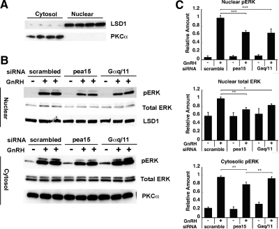
Effect of PEA-15 and Gαq/11 silencing on GnRH-induced ERK activation in the nucleus vs. cytosol. A, Isolation of the nuclear and cytoplasmic fractions of LβT2 cells. Cells were fractionated into nuclear and cytosolic extracts. Aliquots of the nuclear and cytoplasmic fractions were subjected to a Western blot analysis using an LSD1- and a PKCα-specific antibody, respectively. LSD1 is a nuclear marker; PKCα is a cytosolic marker. B, LβT2 cells were serum starved overnight, transfected with scrambled, PEA-15, or Gαq/11 siRNA and stimulated with 100 nm GnRH for 5 min. Cells were fractionated into nuclear and cytoplasmic extracts, and the aliquots of the nuclear and the cytoplasmic fractions were subjected to a Western blot analysis using phospho-ERK and LSD1-specific antibodies (upper blot) or using phospho-ERK- and PKCα-specific antibodies (lower blot). LSD1 was used as a loading control for the upper blot, whereas PKCα was used in the lower blot. C, Quantification of Western blot (B) densitometry, plotted as mean ± sem. Results of triplicate experiments were combined for analysis. The y-axis shows band intensities relative to the positive control (scrambled siRNA, + GnRH). Two-way ANOVA, n = 8; ***, P < 0.005; **, P < 0.01; *, P < 0.05.

