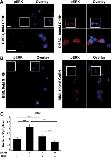
Fig. 4.
GnRH-induced accumulation of phospho-ERK in the nucleus is PKC- dependent. LβT2 cells were serum starved overnight, pretreated with either dimethyl sulfoxide (DMSO) (A) or 10 μm BIMI (B) for 30 min and stimulated or not with 100 nm GnRH for 5 min. Cells were then fixed and stained with an anti-phospho-ERK antibody (red) and DAPI (blue), as indicated in Materials and Methods. The boxed areas are shown at high magnification. Scale bar, 20 μm. C, Single-cell quantification of phospho-ERK fluorescence using the 3D-CatFISH image analysis suite (70, 71). The y-axis displays the nucleus to cytoplasm fluorescence ratio. NS, Nonsignificant. Two-way ANOVA; ***, P < 0.005.

