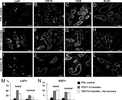
Fig. 6.
Localization of Lef1+ cells in bicalutamide-treated prostates. A–K, Sections of ventral prostates lobes from P30 control-untreated males (A–D), male siblings treated for 7 d with Casodex (30 mg/kg·d in drinking water) starting at P23 (E–H), and male siblings treated with bicalutamide and analyzed after 4 d of recovery (I–L). Sections were immunolabeled for Lef1 (A, E, and I) and nuclei stained for 4′,6-diamidino-2-phenylindole (DAPI) (A); then for CK14 (B, F, and J), CK8 (C, G, and K), and Ki67 (D, H, and L). A, In untreated prostates, Lef1 localized to the basal epithelium (arrowhead). In Casodex-treated prostates (E and I), Lef1 was detected in the basal (arrowheads) and luminal (arrows) epithelium and also in periepithelial mesenchyme (asterisk). Scale bars, 100 μm. M and N, Percentages of the Lef1+ (M) and Ki67+ (N) cells in the control (black bar), treated (stripped bar), and recovering prostates (white bar). P < 0.05.

