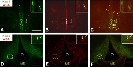
Fig. 7.
LepRb neurons lie in close contact with AVPV and Arc Kiss1 neurons. Representative images of Kiss1-IR (panels A and D, green) and WGA-IR (panels B and E, red) and merged (panels C and F) in the AVPV (panels A–C) and ARC (panels D–F) of a LepRbWGA mouse. Yellow arrows indicate colocalization of Kiss1-IR and WGA-IR. Inset represents digital zoom of the area indicated by dashed box. Scale bars, 200 μm. 3V, Third ventricle; ME, median eminence.

