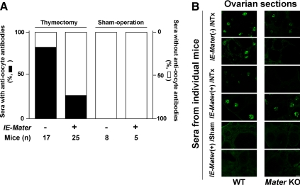
Fig. 3.
Antioocyte autoantibodies in wild-type and IE-Mater transgenic mice. A, Incidence. Based on immunofluorescence on frozen ovarian sections (B) and confirmation with the immunoblotting assay, the sera from 82% of the wild-type (n = 17) and 28% of the IE-Mater transgenic (n = 25) NTx-treated mice contained autoantibodies against oocytes. Thus the incidence of antioocyte antibodies was significantly reduced in the thymectomized IE-Mater transgenic mice (P = 0.01). None of the wild-type (n = 8) and IE-Mater transgenic (n = 5) mice with sham operations produced antioocyte antibodies. B, Representative indirect immunofluorescence. Immune sera from mice with confirmed autoimmune oophoritis are capable of reacting with oocyte proteins other than MATER. Shown are results using sera from NTx-treated mice that had histologically confirmed autoimmune oophoritis: wild-type mice (rows 1 and 2) and IE-Mater transgenic mice (rows 3 and 4). Oocyte autoantibodies were detected using FITC-conjugated goat antimouse IgG as the secondary antibody on wild-type frozen mouse ovarian sections (left column) and Mater knockout (KO) mouse frozen ovarian sections (right column). Results using sera from mice without autoimmune oophoritis are also shown (rows 5 and 6). Negative controls with the secondary antibody alone in both wild-type and Mater null ovarian sections gave little fluorescence (data not shown). Scale bar, 100 μm. WT, Wild type.

