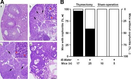
Fig. 4.
Incidence of autoimmune oophoritis after NTx in wild-type and IE-Mater transgenic mice. A, Histopathology of autoimmune oophoritis after NTx. Representative sections of ovarian histology with hematoxylin and eosin staining are shown for sham-operated wild-type mice (a), thymectomized wild-type mice (b), and thymectomized IE-Mater transgenic mice (c and d) at age 10 wk. Lymphocytic infiltration and ovarian follicular destruction indicating autoimmune oophoritis were observed in most of the thymectomized wild-type (b) and some of the thymectomized IE-Mater transgenic (c) mice. Note the lack of primordial follicles in these sections (b and c). Areas of infiltration are indicated with black arrowheads, and insets show the presence of lymphocytes and plasma cells on higher magnification (panels b and c, red arrowheads). However, the ovarian histology of many of the thymectomized IE-Mater transgenic mice (d) appeared normal. Scale bar, 100 μm. B, Incidence of autoimmune oophoritis in mice after NTx. Based on histological evaluations, 94% of the thymectomized wild-type mice (n = 17) and 56% of the thymectomized IE-Mater transgenic mice (n = 25) developed autoimmune oophoritis (solid bars), and thus the incidence of developing autoimmune oophoritis was significantly reduced (P < 0.02). All of the wild-type (n = 10) and IE-Mater transgenic mice (n = 5) with sham operations showed no evidence for oophoritis (open bars).

