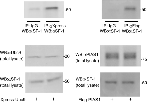
Fig. 1.
Interaction of Ubc9 and PIAS-1 with SF-1 in H295R cells. H295R cells were transfected with Xpress-tagged Ubc9 or Flag-tagged PIAS1. Whole-cell extracts were subjected to IP with anti-Xpress, anti-Flag antibody, or IgG and immunoprecipitates were subsequently analyzed by Western blotting (WB) with anti-SF-1 (top panel). Expression levels of overexpressed Ubc9 or PIAS1 (middle panels) and endogenous SF-1 (bottom panel) in total lysate were analyzed by Western blotting.

