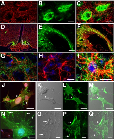
Fig. 1.
SynCAM immunoreactivity in the female mouse hypothalamus (late juvenile period, 28 d of age) is extensively associated with astrocytes and GnRH neurons. A, SynCAM (red). B, GFP-expressing GnRH neurons (green). C, Merged image of single confocal sections. D, Merged confocal image shows that SynCAM (red) is extensively distributed throughout the hypothalamus and is associated with GnRH nerve terminals (green) in the median eminence. E and F, Zoomed confocal images of the area demarcated by the white box in D showing GnRH nerve terminals (E, green) and merged SynCAM/enhanced GFP images (F). G, Merged projection of confocal images showing SynCAM immunoreactivity (green) associated with GFAP-labeled astrocytes (red). H, Hypothalamic tissues incubated with only GFAP antibodies (red). I, Zoomed merged projection of confocal images showing SynCAM immunoreactivity (green) associated with GFAP-labeled astrocytes (red). J, GT1-7 cells stained with GnRH antibodies (red) show intense SynCAM staining (green) in regions of cell contact (arrows). K–M, Projection of confocal images showing GT1-7 adhesion points using DIC (arrow, K) and the corresponding cellular distribution of SynCAM (green, L). The merged image is shown in M. N, Projection of confocal images showing SynCAM immunoreactivity (green, points of abundance denoted by arrows) in cultured hypothalamic astrocytes. The inset in N shows astrocytes stained only with GFAP antibodies (red) and Hoescht. O and P, Projection of confocal images showing astrocyte adhesion points using DIC (arrow, O) and the corresponding cellular distribution of SynCAM (green, P). Q, Merged image of O and P. Bars (A–C), 10 μm; (D), 100 μm; (E and F), 20 μm; (G–J), 10 μm; (K–M), 15 μm; (N), 10 μm; (O and P), 20 μm. Cell nuclei in G–I and N (blue) are stained with Hoescht.

