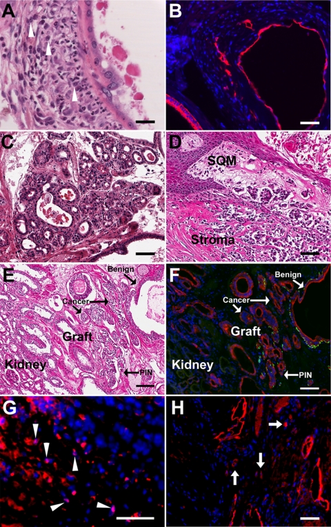
Fig. 7.
Prostate cancer in chimeric prostate renal grafts induced by T+E2 treatment for 2–4 months. A, H&E section of graft exposed to T+E2 for 2 months reveals neoplastic epithelium with enlarged nuclei and prominent nucleoli and their local invasion into the underlying stromal region (cells highlighted with arrowheads). B, CK8/18 immunostained acini shown in A confirms the local invasion of neoplastic cells into the stroma. C and D, Within 2 months of elevated T+E2 exposure, full malignancy was induced in another chimeric renal graft as evidenced by irregular small to medium size glandular structures, abortive glandular lumen, back-to-back lumens, infiltrative glands, and loss of the basement membrane and basal cell layer. Multiple lesions of this type were observed within the graft. E and F, Adjacent sections of a chimeric prostate graft after 4 months of T+E2 treatment with H&E stain (E) and immunofluorescent labeling with CK8/18 (red) and CK14 (green) (F). Staining showed heterogeneous glandular structures with mixture of normal glands, PIN lesions, and carcinoma as well as invasion into kidney. G, Immunostaining with antibody specific to human nuclear antigen (pink nuclei, arrowheads) identifies the human origin of infiltrating epithelial cells (CK8/18+, red) within the stroma. H, Immunolabeling for PSA (red) confirms the human identity of the cancerous ducts and infiltrating cells (arrows) in the grafted tissue. Scale bar, 50 μm.

