Fig. 7.
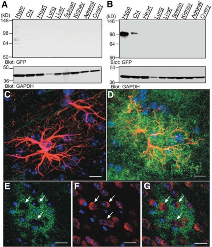
DN SynCAM1 is selectively expressed in astrocytes within the adult female hypothalamus. A, DN SynCAM1 protein, detected with GFP antibodies, is absent in WT tissues. B, DN SynCAM1 protein expression is confined to the central nervous system in transgenic mice. Ctx, Cerebral cortex; Hypo, hypothalamus. C–G, DN SynCAM1 immunoreactivity is confined to astrocytes in the transgenic mouse brain. C, Merged confocal projection images showing the absence of DN SynCAM1 immunostaining (GFP antibodies, green) in WT hypothalamic astrocytes identified with antibodies to GFAP (red). D, DN SynCAM1 is abundant in hypothalamic astrocytes from a DN SynCAM1 animal. E–G, DN SynCAM1 is not expressed in neurons. E, Single confocal section showing DN SynCAM1 immunoreactivity (green) associated with cellular structures surrounding immunonegative cells (identified by Hoechst staining of cell nuclei). F, Immunostaining of neurons using HUC/D antibodies (red). G, Merged image of single confocal sections showing that HUC/D immunoreactive cells are devoid of DN SynCAM1 protein. Bars (C and D), 20 μm; (E–G), 40 μm. Cell nuclei (blue) are stained with Hoechst.

