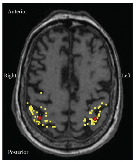
Figure 1.
Motor cortex representation area of a control patient. TMS was focused at the “hotspot” of thenar muscle representation on the primary motor cortex (M1). The yellow dots present stimulation locations during the mapping of the hotspot in the vicinity of M1. The red spots indicate the hotspots, which were located within normal variation in each group [57].

