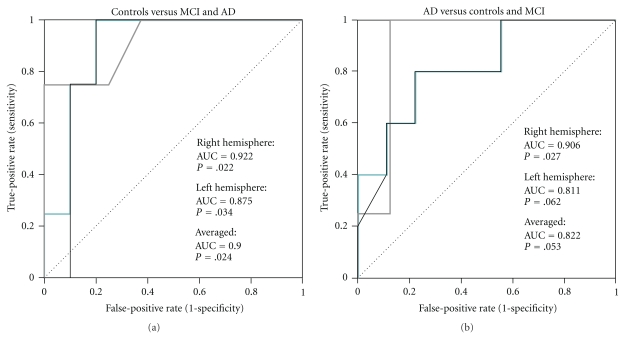
Figure 4.
Receiver operating characteristic (ROC) curves for distinguishing (a) controls from MCI and AD, and (b) and AD patients from MCI and control subjects based on TMS-EEG P30. Turquoise line indicates the ROC curve for averaged data, while the grey and black lines indicate ROC curves for the right and left hemisphere, respectively. The area under the ROC curve (AUC) has been given separately for the averaged (P30mean) and hemispheric data. The asymptotic significance has been indicated with the null hypothesis of AUC = 0.5 (diagonal line).

