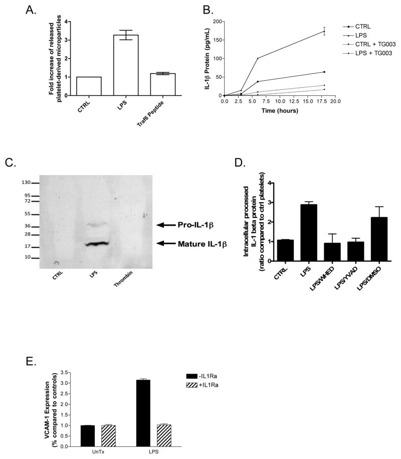
Figure 1.
LPS–stimulated platelets splice IL-1β RNA, process IL-1β protein, and shed IL-1β-laden microparticles, which activate endothelial cells. (A) Platelet microparticles were collected after overnight LPS exposure (100 ng/mL in the presence of recombinant CD14 and LPS-binding protein) or a TRAF6 interacting chimeric peptide before quantitation by flow cytometry. (B) Human platelets (4×10⁸ per condition) were treated with LPS for the stated times in combination with recombinant human CD14 and LPS-binding protein and secreted IL-1β protein was measured by ELISA. (C) Microparticles generated after overnight treatments were resuspended in reducing SDS sample buffer and probed for IL-1β after SDS-PAGE in a western blot. (D) Platelet lysates were probed by ELISA for mature and pro-IL-1β protein after LPS treatment, in the presence or absence of caspase inhibitors. (E) Purified platelet microparticles were added to HUVEC’s for 6 hours before VCAM-1 expression was determined by ELISA. IL-1Ra (150ng/mL) was added 30 minutes prior to microparticle addition. Error bars = +/− 1 SE, N=3 for (A–D) and N=8 for (E).

