Figure 6.

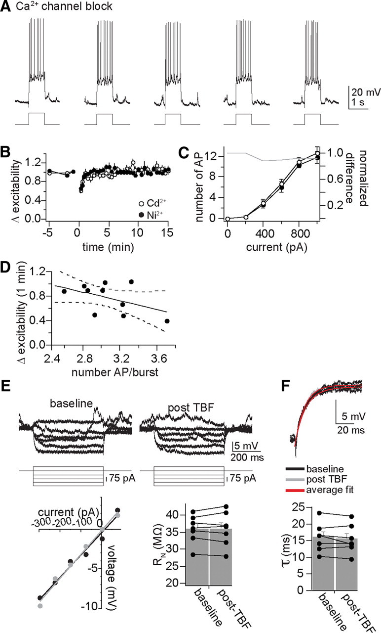
Plasticity of membrane excitability is dependent upon calcium channels. A, With intracellular blockade of Ca2+ channels with Cd2+ (depicted here) or Ni2+, TBF was not able to cause a decrease of membrane excitability. B, Plotted over time, intracellular application of Cd2+ or Ni2+ blocked TBF-induced reductions of membrane excitability. C, Even when membrane excitability was measured over a range of current steps to produce a full input–output curve, TBF did not decrease excitability if Ca2+ channels were blocked (plotted here is intracellular Cd2+; gray line represents the difference between baseline and post-TBF excitability). D, This lack of effectiveness of TBF is not caused by insufficient action potential firing during TBF, as most of the experiments were performed with >2.5 action potentials per burst, and there was still minimal plasticity. E, The effects of TBF on neuronal input resistance were blocked by intracellular blockade of calcium channels with Cd2+ or Ni2+ (displayed in this figure are examples with Cd2+, baseline Rn = 40 MΩ, post-TBF = 42 MΩ; black circle, baseline; gray circle, post-TBF). F, The effects of TBF on membrane time constant were also blocked by intracellular blockade of calcium channels (bottom; baseline, 12.7 ms; post-TBF, 13.1 ms). In almost all control neurons tested (see above), TBF decreased Rn and the time constant, while this effect of TBF was rarely observed if calcium channels were blocked (plotted here with intracellular Cd2+).
