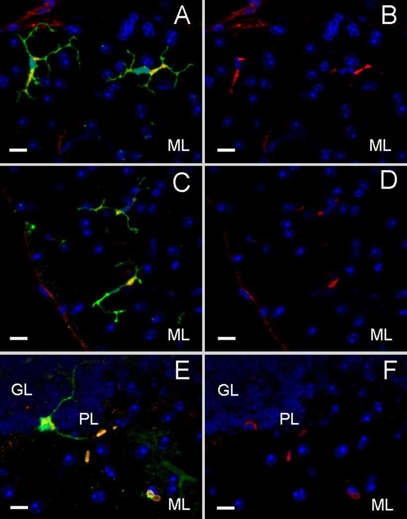
Figure 4. Purkinje heterokaryons and bone marrow-derived microglia express the c-Myc/his reporter tag in vivo.
A) A GFP+ cell type found in Sca1154Q/2Q recipient cerebella has the morphology of a microglial cell (green) and colocalizes with the surface marker CD11b (red) as shown in B. C) 20–30% of the microglia (green) as shown in D) expressed c-Myc (red) in the cytoplasmic regions where both of the modifier genes are localized. E) Fused, GFP+ Purkinje heterokaryons (green) are also immunopositive for c-Myc (red) in F. (scale bar=10µm, GL=inner granule layer, PL=Purkinje layer, ML=molecular layer. DAPI=blue)

