
Figure 5.
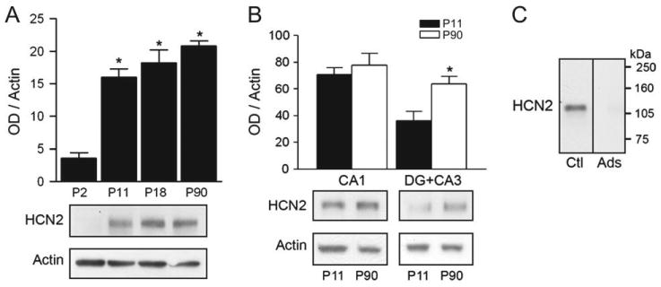
Quantitative western blot analyses of HCN2 protein expression in developing hippocampus. (A) Top: In whole hippocampus, HCN2 protein levels are relatively low on P2, increase between P2 and P11, but not thereafter. Bottom: Representative western blot. Optical density (OD) of HCN2-immunoreactive bands was normalized to that of actin for each lane (*denotes significance when compared with P2, P < 0.05). (B) Top: Regional analyses reveal that HCN2 protein and mRNA expression (see Fig. 4A) are comparable in CA1. However, in DG + CA3, HCN2 protein levels are significantly higher on P90 compared with P11, in contrast to mRNA levels (*denotes significance between groups; t-test: P < 0.05). Bottom: Representative western blots. (C) Immunoreactive-HCN2 bands (Ctl) have an apparent molecular weight of ∼115 kDa. Preadsorption (Ads) with antigen abolishes immunoreactivity. Note that because both HCN1 and HCN2 are detected using the same secondary antibody (anti-rabbit IgG), a control experiment omitting the primary antibody is provided for HCN1 only (Fig. 2C).