
Figure 9.
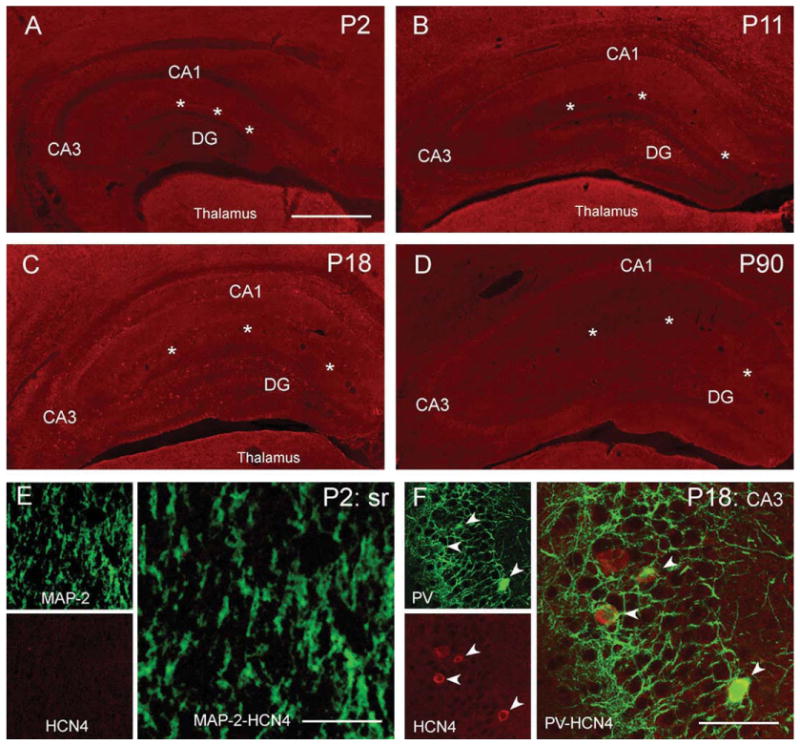
Subcellular localization of HCN4 channels in developing hippocampus. (A–D) Low-magnification photographs show relatively low levels of HCN4 immunoreactivity in hippocampus on P2 (A), P11 (B), P18 (C), whereas HCN4 signal was poorly detectable on P90 (D). On P2, HCN4 signal was distributed over the pyramidal cell layer and weakly apparent in the dendritic fields of CA1 (A); however, dual labeling with MAP-2 did not provide evidence for a dendritic localization of these channels (E). HCN4 expression in interneurons was clearly visible on P18 (C). HCN4-immunoreactive interneurons were mainly associated with the pyramidal cell layer (F, arrowheads) and, similar to HCN1 and HCN2, coexpressed PV (F) but not CCK. In contrast to the situation with HCN1 and HCN2, HCN4 protein expression was found primarily in somata of PV-positive basket cells (F, arrowheads) and rarely in axonal terminals. slm, stratum lacunosum-moleculare; sr, stratum radiatum; asterisks demarcate hippocampal fissure. Virtual slice thickness: (A–D) = 15 μm; (E, F) = 1 μm. Scale bar: (A–D) = 900 μm; (E) = 27 μm (left panels), 13.5 μm (right panel); (F) = 120 μm (left panels), 60 μm (right panel).