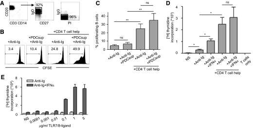
Figure 1. PDCs enhance naïve B cell proliferation induced by T cell help.
(A) Highly enriched CD19+CD27–IgD+IgG– naïve B cells were isolated from PBMCs by magnetic bead isolation. Numbers indicate percentage of gated cells. (B) Anti-Ig and supernatants from TLR7/8-stimulated PDCs (PDCsup) were added to CFSE-labeled, naïve B cells in the presence or absence of T cell help for 6 days. Proliferation was measured by flow cytometry. Numbers indicate percentage of dividing B cells. (C) Compiled data are shown (mean±sd; n≥4 donors). (D) Naïve B cells were stimulated with anti-Ig and human IFN-α in the presence or absence of T cell help, and proliferation was measured by thymidine incorporation (n=3). (E) Naïve B cells were stimulated with increasing concentrations of TLR7/8 ligand in the presence or absence of IFN-α, which is known to increase the sensitivity of B cells to TLR7/8 ligand. Proliferation by thymidine incorporation was measured at Day 5.

