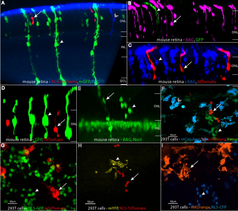
Figure 7.

Retroviruses with distinct histochemical and fluorescent reporters. Combinations of BAG, LIA, and QC viruses were injected in vivo into the subretinal space of P0 retinas, and the retinas were examined at P21. The reporter combinations shown are (A) GFP (anti-GFP, in green, arrowhead), mCherry (anti-RFP, in red, arrow), and BAG (anti-β-galactosidase, in blue, chevron), (B), GFP (anti-GFP, in green, arrowhead) and BAG (anti-β-galactosidase, in magenta, arrow) (C), tdTomato (anti-RFP, in red, arrow) and BAG (anti-β-galactosidase, in blue, arrowhead), (D) GFP (anti-GFP, in green, arrowhead) and tdTomato (anti-RFP, in red, arrow), (E) NinII (anti-β-gal, in green, arrowhead) and BAG (anti-β-gal, in green, arrow) with Chx10-NLS-GFP, a gene in the transgenic background, marking bipolar cell nuclei. The retinal layers are labeled in panels A–E as follows: ONL = outer nuclear layer, INL = inner nuclear layer, GC = ganglion cell layer. Panels F–I show images of infected 293T cells. These combinations are: (F) membrane Cerulean (blue, chevron), Venus (green, arrowhead), and NLS-mCherry (red, arrow), (G) NLS-GFP (green, arrowhead) and membrane tdTomato (red, arrow), (H) NLS-tdTomato (red, arrow) and membrane YFP (yellow, arrowhead), and (I) membrane Kusabira Orange (orange, arrow) and NLS-CFP (blue, arrowhead).
