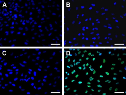
Figure 8.
Apoptotic cells labeled with TUNEL assay in the ARPE-19 cultures. Fluorescence micrographs of control cells (without materials) (A), and cells after exposure to 5 mg of different types of chitosan membranes (B) Chi, (C) GP-chi, and (D) GTA-chi for 24 h at 37 °C. Blue fluorescence is DAPI nuclei staining. Green fluorescence is TUNEL-positive nuclei staining. Scale bars indicate 30 μm.

