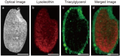
Figure 4.
Ion image of nutrients in rice (Oryza sativa). Scale bar: 1 mm. (a) Optical image of a rice kernel (Hinohikari). (b) Distribution of lysolecithin. (c) Distribution of triacylglycerol. (d) Merged image of lysolecithin (red) and triacylglycerol (green). The rice kernel section can be prepared using adhesive film.

