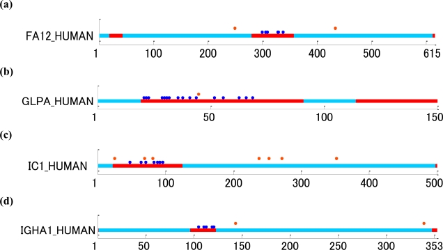
Figure 5.
Glycosylation sites plotted along with the distinction between structural domains and ID regions of human glycoproteins. The light blue and red regions correspond to structural domains and ID regions, respectively, and the blue and orange dots indicate mucin-type O-linked (GalNAc) and N-linked sites, respectively. (a) FA12_HUMAN: coagulation factor XII with O-linked (GalNAc) modifications at T299, T305, S308, T328, T329 and T337, and N-linked (GlcNAc) modifications at N249 and N433. (b) GLPA_HUMAN: glycophorin-A with O-linked sites at S21, T22, T23, T29, S30, T31, S32, T36, S38, S41, T44, T52, T56, S63, S66 and T69, and N-linked site at N45. (c) IC1_HUMAN: plasma protease C1 inhibitor with O-linked sites at T48, S64, T71, T83, T88, T92 and T96, and N-linked sites at N25, N69, N81, N238, N253, N272 and N352. (d) IGHA1_HUMAN: Ig α-1 chain C region with O-linked sites at S105, S111, S113, S119 and S121, and N-linked sites at N144 and N340.

