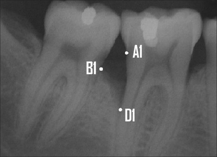
Figure 2.

Landmarks selected: A1- CEJ of the tooth involved in the intrabony defect. B1- The most coronal position of the alveolar bone crest of the intrabony defect when it touches the root surface of the adjacent tooth (the top of the crest). D1- The most apical extension of the intrabony destruction where the periodontal ligament still retained its normal width (the bottom of the defect)
