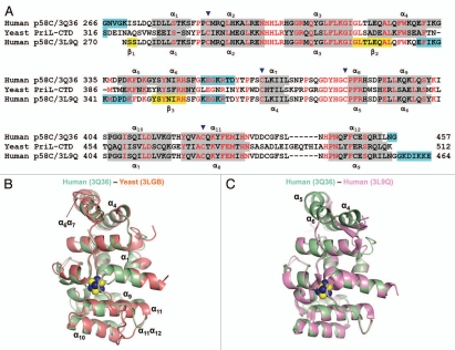
Figure 2.
Comparison of the human p58C/3Q36 structure determined in current work with the corresponding domain structures reported for the yeast and human proteins. (A) Structure-based amino acid sequence alignment of the human and yeast primase 4Fe-4S domains. The sequences corresponding to α-helices and β-strands are highlighted by grey and yellow color, respectively. Conserved residues are shown in red and residues not in the final model are highlighted by cyan color. Residues involved in 4Fe-4S cluster coordination are indicated by triangles. (B and C) Comparison of the human p58C/3Q36 with the structures of the (B) yeast (3LGB) and (B) human (3L9Q) 4Fe-4S cluster domains. The labeled regions exhibiting the significant differences are discussed in the text

