
Figure 3.
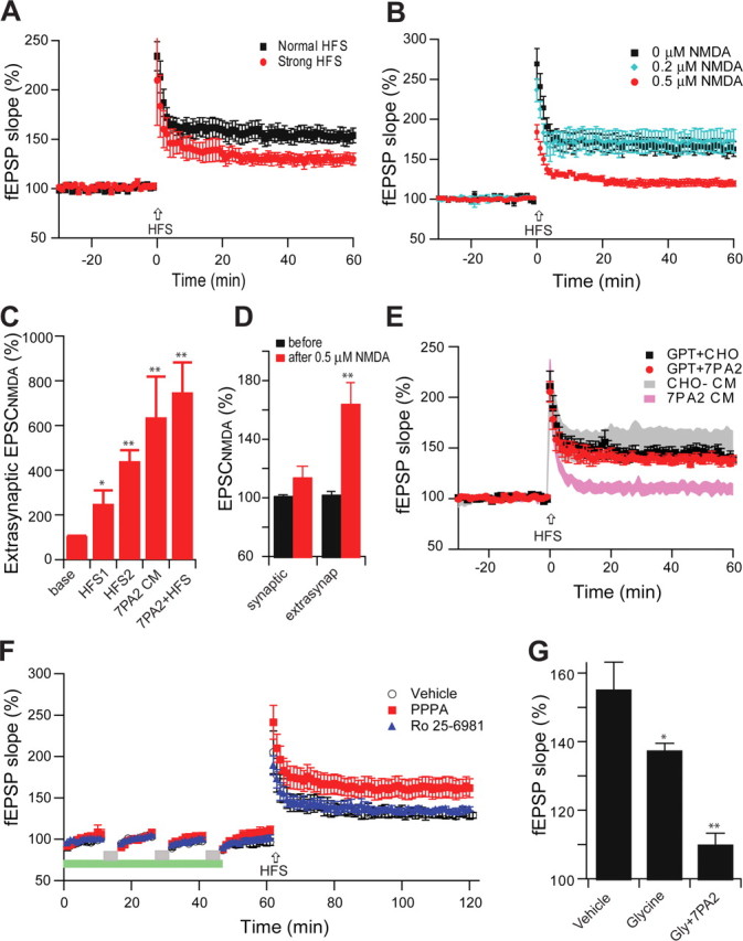
Soluble Aβ impairs LTP via increased extracellular glutamate levels in the Schaffer collateral-CA1 pathway of hippocampus. A, In contrast with the conventional high-frequency stimulation (two trains of 100 Hz, 1 s, separated by 20 s at test stimulus intensity, black squares, n = 6), a strong HFS (at 1.5× test stimulus intensity) significantly reduces the LTP induction (red circles, n = 5). B, A low dose of NMDA (0.5 μm, red circles, n = 7) prevented HFS-induced hippocampal LTP. C, Extrasynaptic NMDA current changes induced by conventional HFS (HFS1), strong HFS (HFS2), 7PA2 CM, or 7PA2 CM plus HFS (7PA2+HFS). D, Responses of isolated NMDAR-mediated synaptic and extrasynaptic (extrasynap) EPSCs to the 0.5 μm NMDA. E, An enzymatic glutamate scavenger system (GPT plus pyruvate) has no significant effect on normal LTP in CHO− CM (black squares, n = 7) but fully prevented the 7PA2 CM impairment of LTP (red circles, n = 8). F, Three repetitive 5 min episodes of subthreshold, low-frequency stimulation (0.5 Hz) (gray bar) desensitize the synaptic NMDAR effect on LTP induction. Green bar represents the time of application of either NR2A-selective (red squares) or NR2B-selective (blue triangles) antagonists given to prevent the desensitization. G, Summary data for the effect of the NMDAR desensitization regulator, glycine (Gly) (100 μm), on Aβ-impaired LTP. Error bars represent ± SEM. *p < 0.05, **p < 0.01.