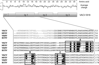
Figure 1.
Sequence analysis of B18 protein. Schematic representation of VACV B18 showing the locations of conserved domains and a plot of charge density along the protein. SP, signal peptide; Ig, immunoglobulin domain. Partial amino acid alignment of the B18 protein (first 171 aa) and its MPXV, VARV, and ECTV orthologues is shown. Boxed regions A–C indicate potential GAG-binding domains identified. Black boxes highlight R and K residues.

