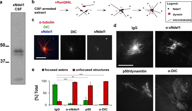
Figure 1.
Ndel1 function is required for microtubule aster formation. (a) Affinity-purified α-Ndel1 antibodies recognize a 39-kD band in Xenopus egg extracts. (b) Schematic of putative role of dynein and Ndel1 in Ran aster formation in egg extracts. (c) Ndel1 colocalizes with dynein in the central focus of asters. (d) Addition of p50/dynamitin, α-dynein intermediate chain (DIC) or α-Ndel1 antibodies disrupts formation of microtubule asters. (e) Quantification of aster formation in the experiment described in panel d. Over 100 microtubule structures were quantified in three independent antibody addition experiments; asterisks indicate statistically significant differences (Student’s t test with P < 0.05; ** indicates P < 0.005; *** indicates P < 0.0005). Bar, 20 µm.

