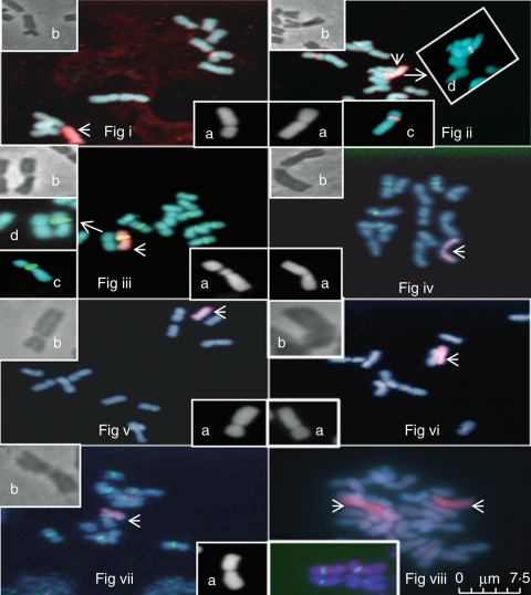
Fig. 1.
Mitosis in the seven monosomic substitution lines. (Fig i–vii) Monosomic lines 1–7; the substituted chromosome is shown by an arrow. Inset images: (a) 4′,6-diamidino-2-phenylindole (DAPI)-stained image of the relevant substitution line; (b) phase contrast image of the relevant substitution line. (Fig ii) Inset images: (c) and d) show the 5s site in monosomic line 2. (Fig iii) Inset images: (c) and (d) show monosomic line 3 carrying the pta71 ribosomal site. (Fig viii) A colchicine doubled line of monosomic line 3 (2n = 28) carrying a disomic substitution. Inset image: (a) shows the disomic chromosome substitutions for monosomic line 3 carrying the two pta71 sites. Scale bar = 7·5 µm.

