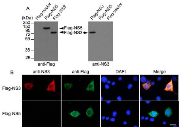
Figure 2.
Analysis of the specificity of the anti-NS3 antibody produced. (A) Western blot analysis of Flag-NS3 expressed in Neuro-2a cells. Cell lysates (20 μg) from Neuro-2a cells transfected with plasmid expressing Flag-NS3 were probed with the anti-NS3 antibody (anti-NS3 panel) and then re-probed with anti-Flag antibody (M2, Sigma) (anti-Flag panel). The plasmid expressing Flag-tagged JEV NS5 protein (Flag-NS5) and Flag-empty vector (Flag-vector) were used as controls. (B) Immunofluorescence analysis of Flag-NS3 expressed in Neuro-2a cells. Neuro-2a cells transfected with plasmid expressing Flag-NS3 were double-immunostained with the anti-NS3 antibody (anti-NS3 panel, red) and anti-Flag antibody (anti-Flag panel, green). The cells transfected with plasmid expressing Flag-tagged JEV NS5 protein (Flag-NS5) were used as controls. The cells were also stained for DNA with 4', 6'-diamidino-2-phenylindole (DAPI) (DAPI panel, blue). Merge panel shows the superimposed image. Bar, 20 μm.

