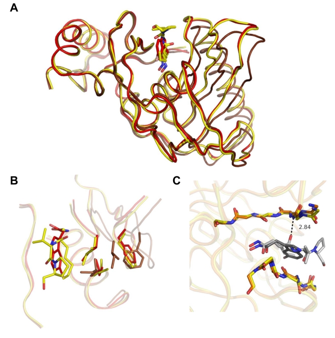
Figure 6. Effect of 6b binding on the conformation of key residues of PDF.
Superimposition of free, 6b-, and actinonin-bound AtPDF indicated in brown, red, and yellow, respectively. (A) Molecule A in the three models was superimposed, resulting in an r.m.s.d. of 0.9 Å for 100% of the Cα. Actinonin is shown in yellow and 6b in red. (B) Conformation of key residues Ile42, Phe58, and Ile130 in the different complexes and in unbound WT AtPDF. Actinonin is shown in yellow and 6b in red. (C) A detailed view of the AtPDF ligand-binding site for both actinonin and 6b complexes, which are indicated by sticks and are superimposed. The two ligands are colored in pale and dark grey, respectively. The hydrogen bond made by actinonin only is shown.

